

Soldiers who died in battle centuries ago will never know that their wounds would one day educate people across the world through digital wizardry.
Skulls and bones from other parts of the body bearing evidence of often horrific injury feature among the University of Bradford’s vast collection of human remains - the largest of any university archaeology department in the country, with more than 4000 skeletons ranging from the Neolithic to the 19th century.
Specimens from the collection, which suffered horrific wounds to the head in battle helped to inform the team examining the fatal combat injury from the body identified as King Richard lll, recently reburied in Leicester.
And now they are helping experts and students from all corners of the globe, through a special online digital resource of human remains.
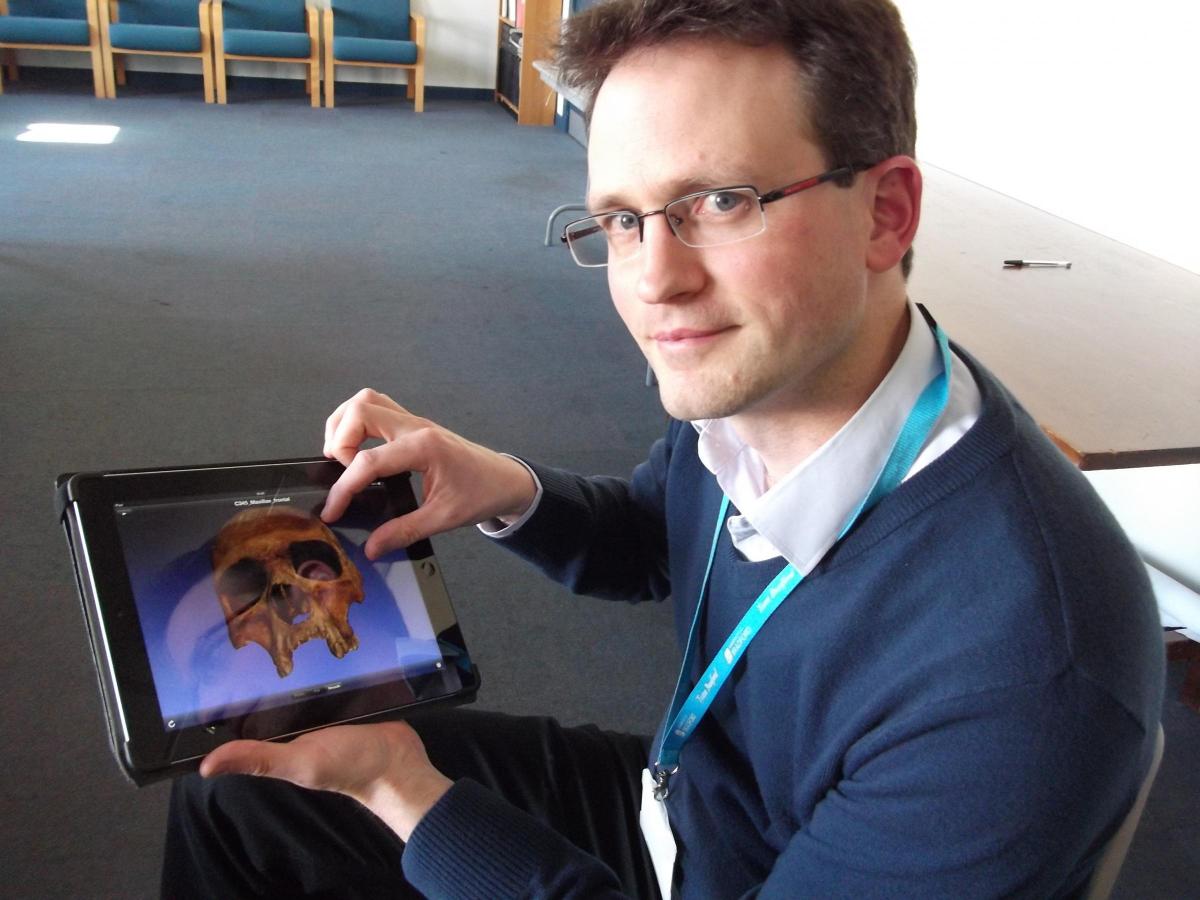
The Department of Archaeological Sciences’ Digitised Diseases project uses the latest 3D laser scanning techniques, CT scans and high resolution photography to create pin-sharp images of human bones which can be viewed at all angles. Together with clinical descriptions and historical illustrations they create an easily accessible archive that can be called up on a laptop, ipad, mobile phone or other electronic device.
The unique resource holds more than 1,620 specimens covering more than 90 chronic pathological conditions affecting adults and children, including common degenerative joint diseases such as osteoarthritis to infections like tuberculosis and metabolic deficiencies such as scurvy and rickets. More than 450 historic images of patients with leprosy - taken in India, Nepal and Ethiopia - have also been digitised.
It forms a unique educational tool that will aid many people, inclduing clinicians, medical students, medical historians, archaeologists, osteologists and palaeopathologists as well as enriching the public’s understanding of anatomy and medical science.
“We always focused upon the project as a learning resource,” says the project’s lead researcher Dr Andrew Wilson. “The really important aspect was focusing on type specimens, with a description of the changes and a clinical synopsis.”
The skeleton collections used in the project - which was Launched at the Royal College of Surgeons in London - are not only those of the university, but from internationally renowned collections from museums and trusts across the country including the Museum of London Archaeology( MOLA), the Hunterian Museum, the Wellcome Museum of Anatomy and Pathology at the Royal College of Surgeons in London. The project presents an opportunity to view collections that have restricted access and are therefore usually only seen by students and researchers.
Each image is accompanied by information about the individual and how the injury came about, if known, for example injuries caused in different ways - accidental, intentional or surgical.
The archaeology of disease was pioneered in Bradford by former GP, Professor Keith Manchester, who still lectures at the university. The institution is now a world leader in the field utilised by experts from across the globe. A team examining the body of King Richard lll visited to compare the wounds from the individual from Greyfriars with the skeletons that were found in a mass grave on the Towton battlefield in 1996. “It helped them to reach a conclusion,” says Andrew.
“We have digitised some of Towton’s specimens to show trauma,” he adds.
Collections of ancient bones invariably include some remains that cannot be handled due to their fragility or could suffer damage from repeated handling. Accessing digitised images allows studies of such remains.
Initially funded by the Joint Information Systems committee (JISC), a body sponsored by Government departments and funding councils, which supports post-16 education and research with a focus on information and communications technology. the project began as a pilot, to bring together the university’s collection, with expertise in visual computing.
Advances in medicine now mean that diseases that would once be left to progress are now treated. Says Dr Cathy Batt**”people will not always see the effects of these diseases. Digitised Diseases allows people to see what would happen if they are left untreated.”
Work has been carried out with other associate partners including the York Bones Forum, who approved the scanning of specimens from various collections including that of the Yorkshire Museum and National Museums Scotland..
“We see it very much as legacy project,” says Andrew, who stresses that many people have been involved in its success. “One that is important not just for Bradford but because of the uptake on an international scale. It is demonstrating its worth and value by other people consulting it and seeing it as a very useful and valuable resource.”
He adds: “We are adding to it constantly. One of our students is now looking at possibly involving groupings of bones.”
digitiseddiseases.wordpress.com;facebook.com


Comments: Our rules
We want our comments to be a lively and valuable part of our community - a place where readers can debate and engage with the most important local issues. The ability to comment on our stories is a privilege, not a right, however, and that privilege may be withdrawn if it is abused or misused.
Please report any comments that break our rules.
Read the rules here